Imagery platform
Observe and understand the infinitely small to model and optimize the industrial process
The imagery platform is equipped with a variety of equipments allowing the acquisition of 2D, 3D and 4D high-resolution images. Obtaining an image allows on the one hand to observe, and on the other hand to follow the evolution of a process when the acquisition is prolonged in time. Beyond imaging, the platform affords a digital post-processing offer allowing to diagnose, analyze quantitatively and model from a real morphology. The platform is commonly used for the observation of biological phenomena and the morphological characterization of biosourced materials, and is open to other observations.
Contact
Aya Zoghlami
aya.zoghlami@centralesupelec.fr
Our equipments
- X-ray nano-tomograph equipped with a 160 kV nano-focus tube and a 150 kV micro-focus tube (EasyTom XL 150-160, RX Solutions) - In situ and surface 3D observation, physically non-destructive with 3D image analysis up to a resolution of 400 nm
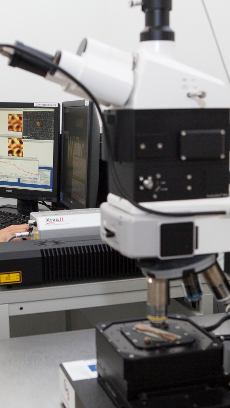
- Confocal Raman microscope equipped with 2 lasers 532 nm, 785 nm and the "TrueSurface" coupling surface analysis and surface topography (Alpha 300R, WITec) - 3D observation of the surface with Raman spectra for chemical analysis
- Apotome microscope with Light Structured Epifluorescence (Zeiss Axiozoom V16, Zeiss) - Creation of optical sections of fluorescent samples
Examples of applications
- Non-destructive 3D imaging of porous composite materials
- 3D image analysis: pore size, fiber orientation, connectivity, volume fractions, etc.
- Observation of materials under stress (mechanical, hydric): measurement, model validation, etc.
- 3D topography of optical sections of fluorescent samples
- Observation of the growth of immobilized mixed crops
- Observation of the growth of filamentous fungi on lignocellulosic substrates
- Characterization of chemical species present in biomass samples